| 1 |
|
2. Fluorescent probes |
|
|
ง
|
7 |
-.50
-.25
+.25
เต็ม
0
-35%
+30%
+35%
|
| 2 |
|
5. Low birthweight for gestation |
|
เนื่องจากความถี่ในช่วงนั้นมีจำนวนมากกกว่าในช่วงอื่นๆ
|
Low birth weight (LBW) is defined as a birth weight of less than 2500 g (up to and including 2499 g), as per the World Health Organization (WHO) [1]. This definition of LBW has been in existence for many decades.
|
7 |
-.50
-.25
+.25
เต็ม
0
-35%
+30%
+35%
|
| 3 |

|
2. Hydrogene |
|
|
|
7 |
-.50
-.25
+.25
เต็ม
0
-35%
+30%
+35%
|
| 4 |
|
5. X-ray crystallography |
|
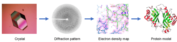
X-ray crystallography is a tool used for determining the atomic and molecular structure of a crystal. The underlying principle is that the crystalline atoms cause a beam of X-rays to diffract into many specific directions (Fig. 2.10). By measuring the angles and intensities of these diffracted beams, a crystallographer can produce a 3D picture of the density of electrons within the crystal.
|
พอได้ผลึก (crystals) ของ protein แล้ว เราก็เอามันไปยิงด้วย x-ray ยิ่งแสง x-ray มีความยาวคลื่นสั้นเท่าไหร่ resolution power ในการที่มองเห็น protein ยิ่งดีขึ้นเท่านั้น และนั่นคือสาเหตุว่าทำไม เราถึงต้องการแสง Synchrotron เพื่อการณ์นี้
เมื่อ protein ถูกยิงด้วยแสง x-ray สิ่งที่เกิดขึ้นคือ แสง x-ray จะไปชนเข้ากับ electrons บนที่อยู่โมเลกุลของโปรตีน ทำให้แสง x-ray เกิดการกระเจิงหักเหแตกต่างกันไปตามมุมและระนาบที่ตกกระทบบน electron การที่แสงกระเจิงออกเป็นหลายๆแสงดังนี้ ก็จะทำให้เกิดโอกาสที่แสงจะรวมตัวกัน หรือ จะทำลายล้างกันไปก็ได้ หากมันเกิดการรวมตัวกัน มันก็จะได้เป็นแสงที่มี intensity สูงมากพอและวิ่งผ่านไปตกกระทบลงบน detector ที่วางไว้หลัง crystal (ดูรูปที่ 1) ทำให้เกิดเป็นจุดสีดำบนจอ detector แต่ละจุดก็คือ intensity ของแสงที่เป็นผลลัพธ์จากแสงย่อยๆร่วมตัวกันนั่นเอง จุดหลายๆจุดที่เห็นบน detector นั่นเราเรียกรวมกันว่า diffraction pattern นั่นเอง และสำหรับแสงที่มันหักล้างกันไป ก็จะหายไปและไปไม่ถึง detector
|
7 |
-.50
-.25
+.25
เต็ม
0
-35%
+30%
+35%
|
| 5 |
ข้อใดไม่เกี่ยวข้องกับ Etiology of Cancer
|
4. Circulation |
|
การไหลเวียนมีความสัมพันธ์โดยตรงกับสาเหตุของโรคมะเร็งน้อยกว่าเมื่อเปรียบเทียบกับทางเลือกอื่นๆ สาเหตุของโรคมะเร็งมักเกี่ยวข้องกับปัจจัยต่างๆ เช่น การพัฒนาอุตสาหกรรม การสัมผัสสิ่งแวดล้อม องค์ประกอบวิถีชีวิตทางวัฒนธรรม และแนวทางปฏิบัติทางการเกษตร ในขณะที่การไหลเวียนจะสัมพันธ์กับการไหลเวียนของเลือดและการเคลื่อนไหวภายในร่างกายมากกว่า ซึ่งไม่ใช่สาเหตุโดยตรงของการพัฒนาของมะเร็ง
|
|
7 |
-.50
-.25
+.25
เต็ม
0
-35%
+30%
+35%
|
| 6 |
ข้อใดไม่เกี่ยวข้องกับหลักการของ blood flow จาก fluid dynamics
|
2. Tissue engineering |
|
|
|
7 |
-.50
-.25
+.25
เต็ม
0
-35%
+30%
+35%
|
| 7 |
ข้อใดเกี่ยวข้องกับหลักการของ Laser physic ที่ใช้ในดวงตา
|
5. ถูกมากกว่า 1 ข้อ |
|
|
|
7 |
-.50
-.25
+.25
เต็ม
0
-35%
+30%
+35%
|
| 8 |
ข้อใดเกี่ยวข้องกับ Peptide Ranker
|
4. Bioactivity potential scoring |
|
Peptide Ranker is often associated with assessing the bioactivity potential of peptides. It is used to predict and score the potential bioactivity of peptides based on various factors, helping in the identification and prioritization of peptides for specific functions or activities.
|
In silico and in vitro methods are useful tools for selection of enzymes to generate bioactive peptides from protein containing biomass. Moreover, they are useful to determine potential bioactivities of peptides prior to chemical synthesis and can save time and money prior to animal studies to determine potential health benefits. A combination of these methods was used previously to identify and confirm the bioactivity of peptides derived from blood proteins [51] and microalgae previously [54]. However, limitations of this approach exist and specifically include limits concerning the folding of protein, which has an impact on how enzymes cut the protein and which in turn can impact production of the resulting peptides. One of the main barriers for entering the human functional foods market is unknown and unstable peptide product qualities. It is required to have analytical methods for characterising the peptide fraction. Today, research groups are using Fourier-transform Infrared (FTIR) fingerprints to gain new insight in quality variations of peptide products. These fingerprints can be related to raw material composition and processing factors [55]. The method used in this study has advantages over in vitro only methods as it can help to predict the best enzymes to use to generate bioactive peptide containing hydrolysates and additionally can predict the most bioactive peptides and those that may be toxic before any in vitro assays are performed.
Two novel ACE-1 inhibitory peptides with amino acid sequences corresponding to IGNNPAKGGLF and YIGNNPAKGGLF were identified from a 3 kDa permeate of a protein hydrolysate generated from the brown seaweed L. digitata. In silico methods also predicted the potential of this seaweed as a source of novel, bioactive peptides that may impart additional health benefits to the consumer including prevention of T2D and antimicrobial activities following GI digestion. Identified, chemically synthesized peptides had ACE-1 inhibition IC50 values of 174.4 µg/mL (0.161 µM) for peptide IGNNPAKGGLF and 133.1 µg/mL (0.107 µM) for peptide YIGNNPAKGGLF and both peptides were similar in terms of bioactivity to other ACE-1 inhibitory peptides identified from tuna and meat muscle previously. This study highlights the potential bioactivity of this brown seaweed. However, future work is required to confirm an anti-hypertensive effect of the seaweed hydrolysate and synthesized peptides in vivo. This work will involve assessment of the L. digitata hydrolysate and synthesized peptides in spontaneously hypertensive rats (SHRs) to assess if the ACE-1 inhibitory peptides have an anti-hypertensive effect in vivo.
|
7 |
-.50
-.25
+.25
เต็ม
0
-35%
+30%
+35%
|
| 9 |
ข้อใดเกี่ยวข้องกับ Ultrasonic Therapy
|
5. ถูกทุกข้อ |
|
Ultrasonic Therapy สามารถ เพื่อรักษาอาการบาดเจ็บของเนื้อเยื่อ และข้อต่อในชั้นลึก
เพื่อลดอาการปวด
เพื่อเพิ่มความยืดหยุ่นให้กับเนื้อเยื่อเกี่ยวพัน
เพื่อลดอาการเกร็งตัวของเนื้อเยื่อและพังผืด
เพื่อลดอาการบวมเนื่องมาจากการอักเสบในระยะแรก
|
อัลตราซาวน์เป็นเครื่องรักษาด้วยคลื่นเสียงความถี่สูง ที่ให้ผลความร้อนในลักษณะความร้อนลึกโดยปล่อยความร้อนลึดออกมาใต้ผิวหนังที่ 2-5 cm. ใช้ลดอาการปวด ลดการอักเสบของเนื้อเยื่อ เพิ่มความยืดหยุ่นของข้อต่อในชั้นลึก ลดอาการบวม และช่วยเร่งการซ่อมแซมเนื้อเยื่อ รวมทั้งคลายการเกร็งตัวของกล้ามเนื้อ
คลื่นอัลตราซาวน์ที่ใช้ในทางก่ายภาพบำบัดมีความถี่อยู่ที่ 0.75 – 3 MHz และมีความเข้มอยู่ในช่วง 0.125 – 3 W/cm²
รูปแบบของคลื่นอัลตราซาวน์
Continuous modeคือการที่คลื่นออกมาตลอดเวลาการรักษา โดยจะให้ผลการรักษาของ Thermal effectคือ มีผลในการลดการยึดแข็งของข้อต่อ ลดการเกร็งของกล้ามเนื้อ และลดการปวด
Pulsed modeคือคลื่นออกมาเป็นช่วงๆ โดยมีช่วงที่คลื่นออก และช่วงคลื่นหยุดพักสลับกันไปตลอดระยะเวลาการรักษา โดยจะให้ผลการรักษาของ Non-Thermal effectคือ มีผลในการเร่งขบวนการสร้างเนื้อเยื่อ กระตุ้นการสังเคราะห์โปรตีน และเร่งการสมานของเนื้อกระดูก
ผลที่ได้จากการรักษาด้วยเครื่องอัลตราซาวน์ทางกายภาพบำบัด
เพื่อรักษาอาการบาดเจ็บของเนื้อเยื่อ และข้อต่อในชั้นลึก
เพื่อลดอาการปวด
เพื่อเพิ่มความยืดหยุ่นให้กับเนื้อเยื่อเกี่ยวพัน
เพื่อลดอาการเกร็งตัวของเนื้อเยื่อและพังผืด
เพื่อลดอาการบวมเนื่องมาจากการอักเสบในระยะแรก/https://www.rehabcareclinic.com/%E0%B9%80%E0%B8%84%E0%B8%A3%E0%B8%B7%E0%B9%88%E0%B8%AD%E0%B8%87%E0%B8%AD%E0%B8%B1%E0%B8%A5%E0%B8%95%E0%B8%A3%E0%B8%B2%E0%B8%8B%E0%B8%B2%E0%B8%A7%E0%B8%99%E0%B9%8C-ultrasound-therapyอ้างิ
|
7 |
-.50
-.25
+.25
เต็ม
0
-35%
+30%
+35%
|
| 10 |
ข้อใดไม่เกี่ยวข้องกับ biophysic ที่ใช้ในการรักษาหัวเข่า
|
2. Propagation speed |
|
Propagation speed, while a relevant parameter in the study of waves, may not be directly associated with the specific application of biophysics used in treating the knee. The other options—Frequency, Pulsed Ultrasound, and Waves' Interaction With Tissue—are more closely related to the principles and techniques employed in biophysics for knee treatment.
|
The propagation speed of sound waves through tissue is an important element of ultrasound scans. Ultrasound machines assume sound waves travel at a speed of 1540 m/sec through tissue 1. In reality, the speed of sound is affected by the density and elasticity of the medium through which it is traveling and these factors are not constant for human tissues. The propagation speed of sound is higher in tissues with increased stiffness and reduced density2.
Examples of propagation velocities in different tissues are given below 2 :
air: 330 m/sec
fat: 1450 m/sec
water: 1480 m/sec
liver: 1550 m/sec
kidney: 1560 m/sec
blood: 1570 m/sec
muscle: 1580 m/sec
bone: 4080 m/sec
Ultrasound machines use echo-ranging to determine the distance between the transducer and reflective interfaces 2. As the machine assumes a constant propagation speed of 1540 m/sec, speed displacement artifact can occur if the sound wave traverses tissues of differing propagation speeds
|
7 |
-.50
-.25
+.25
เต็ม
0
-35%
+30%
+35%
|
| 11 |
ข้อใดเกี่ยวข้องกับ The Scientific Status of the Linear Hypothesis and Radiobiological Data
|
1. Hypothetical Harm |
|
The concept of a practical threshold is often discussed in the context of the scientific status of the linear hypothesis and radiobiological data, particularly in the field of radiation biology. The idea is whether there is a dose threshold below which no harmful effects are observed, challenging the linear no-threshold hypothesis.
|
Feature engineering and polynomial regression
The linear hypothesis in Eq. (15.1) can easily be extended to capture more complex, nonlinear problems through the addition of nonlinear features or feature combinations. For example, for a two-dimensional (2D) input , we can define new features or and add these to our hypothesis. The manual process of creating new features is called feature engineering and ideally involves previous domain knowledge about the problem at hand. A common, systematic way of increasing model complexity are polynomial features, which use polynomial combinations of the features with degree less than or equal to the specified degree. For example, for a 2D input , the two-degree polynomial features are , resulting in the hypothesis/ อ้างอิง https://www.sciencedirect.com/topics/engineering/linear-hypothesis
|
7 |
-.50
-.25
+.25
เต็ม
0
-35%
+30%
+35%
|
| 12 |
ข้อใดไม่เกี่ยวข้องกับ optical imaging ในทางการแพทย์
|
1. Magnetic Resonance Imaging |
|
Optical imaging in the medical context involves techniques that use visible light or near-infrared light, such as fluorescence imaging. Magnetic Resonance Imaging (MRI) primarily relies on the principles of nuclear magnetic resonance and is not directly associated with optical imaging methods.
|
We imaged a phantom (Fig. 2a) consisting of a bottle filled with ethylene glycol (purity 99.8%; natural abundance, 1.1%, 13C; triplet with JCH of 142 Hz). The recorded image (Fig. 2c) shows the spatial density of 13C atoms in a cross-section through the bottle. As we expected, the image does not have a uniform signal-to-noise throughout the volume because we used a surface coil where the detected signal decays with increased distance to the coil (Fig. 2b). This effect is not corrected in the image processing.
To each image voxel corresponds a spectrum obtained from the spin-precession (Fig. 2d). For ethylene glycol, that spectrum has a characteristic triplet as shown in the reference measurement (Fig. 2d), obtained with a commercial coil but still using the same phantom and scanner. The spectrum obtained with the transducer (Fig. 2e) shows the same triplet. Additionally, the two detection schemes have different background noise; for the standard electronic amplifier the noise is flat in a broad window, but for the transducer it has a distinctive narrow Lorentzian lineshape plus an offset (Fig. 2e).
We believe the Lorentzian feature comes from the membrane’s spectral response because the Lorentzian peak frequency and linewidth changed with the bias power as we expect from the electromechanical interaction13,18. Additionally, moving the bias frequency also shifted the detected signal with respect to the noise peak and scaled its amplitude following the peak shape. This is an expected behaviour if the MR signal drives the membrane’s motion and therefore further corroborates that the spectral shape of the noise is related to the mechanical motion. Furthermore, the transducer’s flat noise background scales with optical power which suggests that it originates from amplitude noise of the laser, most likely due to shot-noise of light. Finally, we have not found any other spectral feature in the circuit response, nor any spurious noise in the setup, that is consistent with the observed Lorentzian peak./อ้างอิง https://www.nature.com/articles/s41598-019-54200-3
|
7 |
-.50
-.25
+.25
เต็ม
0
-35%
+30%
+35%
|
| 13 |
ข้อใดเกี่ยวข้องกับ cryo-ET on FIB-fabricated lamellae
|
2. cellular morphology at nanometer resolution |
|
Cryo-electron tomography (cryo-ET) on focused ion beam (FIB)-fabricated lamellae is a technique used to study cellular morphology at a very high resolution, typically in the nanometer range.
|
Cryogenic electron microscopy and data processing enable the determination of structures of isolated macromolecules to near-atomic resolution. However, these data do not provide structural information in the cellular environment where macromolecules perform their native functions, and vital molecular interactions can be lost during the isolation process. Cryogenic focused ion beam (FIB) fabrication generates thin lamellae of cellular samples and tissues, enabling structural studies on the near-native cellular interior and its surroundings by cryogenic electron tomography (cryo-ET). Cellular cryo-ET benefits from the technological developments in electron microscopes, detectors and data processing, and more in situ structures are being obtained and at increasingly higher resolution. In this Review, we discuss recent studies employing cryo-ET on FIB-generated lamellae and the technological developments in ultrarapid sample freezing, FIB fabrication of lamellae, tomography, data processing and correlative light and electron microscopy that have enabled these studies. Finally, we explore the future of cryo-ET in terms of both methods development and biological application./อ้างอิง https://www.nature.com/articles/s41592-023-01783-5
|
7 |
-.50
-.25
+.25
เต็ม
0
-35%
+30%
+35%
|
| 14 |
Method ใดควรนำมาเพื่อใช้ในการวัด Creatinine
|
2. HPLC |
|
HPLC หรือ High-Performance Liquid Chromatography เป็นเทคนิคทางวิทยาศาสตร์ที่บ่งชี้ถึงการวัดปริมาณสารต่างๆ ในตัวอย่าง และเป็นวิธีที่ใช้กันอย่างแพร่หลายในการวัด Creatinine ในทางทางการแพทย์และทางวิทยาศาสตร์ทางคลินิก.
|
Creatine is produced by the body and plays a major role in recycling ATP. It also stabilizes pH in certain tissues. Creatine can be analyzed by this reverse phase (RP) HPLC method with simple conditions. The mobile phase contains acetonitrile (MeCN), water, and TFA. For Mass-Spec (MS) compatible applications the phosphoric acid needs to be replaced with formic acid. Smaller 3 µm particle columns are available for fast UPLC applications. This liquid chromatography method is scalable and can be used for isolation of impurities in preparative separation. It also suitable for pharmacokinetics./อ้างอิง https://sielc.com/Application-HPLC-Separation-of-Creatine-Creatinine-and-Histidine
|
7 |
-.50
-.25
+.25
เต็ม
0
-35%
+30%
+35%
|
| 15 |
What techniques used to provide valuable information in deciphering functional mechanisms of proteins/peptides
|
1. Molecular dynamics simulations |
|
can provide valuable information in deciphering functional mechanisms of proteins/peptides and other biomolecules, overcoming the rigid sampling limitations in docking analysis.
|
In silico tools, such as molecular docking, are widely applied to study interactions and binding affinity of biological activity of proteins and peptides. However, restricted sampling of both ligand and receptor conformations and use of approximated scoring functions can produce results that do not correlate with actual experimental binding affinities. Molecular dynamics simulations (MDS) can provide valuable information in deciphering functional mechanisms of proteins/peptides and other biomolecules, overcoming the rigid sampling limitations in docking analysis. This review will discuss the information related to the traditional use of in silico models, such as molecular docking, and its application for studying food proteins and bioactive peptides, followed by an in-depth introduction to the theory of MDS and description of why these molecular simulation techniques are important in the theoretical prediction of structural and functional dynamics of food proteins and bioactive peptides. Applications, limitations, and future prospects of MDS will also be discussed./อ้างอิง https://pubmed.ncbi.nlm.nih.gov/34990125/
|
7 |
-.50
-.25
+.25
เต็ม
0
-35%
+30%
+35%
|
| 16 |
สารละลาย 0.30 M solution of NaCl is equivalent to 0.60 OsM.
|
3. 0.8 Osm. |
|
The osmolality (OsM) is a measure of the concentration of solute particles per unit of solvent, and in this case, a 0.30 M solution of NaCl is equivalent to 0.60 OsM.
|
Normal saline หรือ 0.9%NaCl solution ถือว่าเป็นสารน้ำที่มีการใช้งานมากที่สุดในวงการแพทย์มาช้านาน แต่ทราบกันหรือไม่ครับ ว่าทำไมมันถึงจัดเป็นสารละลายชนิด Isotonic
ยิ่งไปกว่านั้น ถ้าเราคำนวณความเข้มข้นของสารละลาย (Osmolarity) จะพบว่า ความเข้มข้นควรจะเป็น 308 mOsm/L ซึ่งสูงกว่า Plasma แล้วทำไมมันยังถึงจัดเป็นสารละลาย Isotonic ล่ะ?
ก่อนอื่นเราต้องทราบถึงความหมาย และที่มาของสารละลายชนิดนี้กันก่อนครับ
👍 0.9% NaCl solution หมายถึง สารละลาย NaCl ที่มีความเข้มข้นของ NaCl (ตัวถูกละลาย) 0.9% โดยมวล ต่อปริมาตรของสารละลาย
👍 0.9% NaCl solution จึงหมายถึง
สารละลายทั้งหมด 100 ml มี NaCl ละลายอยู่ 0.9g
หรือถ้าเทียบสารละลาย 1 L ก็จะมี NaCl = 0.9 x 10 = 9 g
9 g ของ NaCl คิดเป็น mol โดยการเอา มวล (หน่วยกรัม) หาร มวลโมเลกุลของ NaCl (NaCl มี molecular weight = 23 + 35.5 = 58.5)
9 g ของ NaCl จึงคิดเป็น = 9/58.5 = 0.154 mol หรือก็คือ 154 mmol
สรุปในสารละลาย 0.9%NaCl 1 L จะมี NaCl ละลายอยู่ 154 mmol
หรือคิดเป็นความเข้มข้นคือ = 154 mmol/L หรือหน่วย Molar นั่นเอง
👍 แต่ NaCl จำนวน 154 mmol/L ที่ละลายอยู่ จะแตกตัวให้ Na+ และ Cl- ดังสมการ
NaCl ↔ Na(+) + Cl(-)
ดังนั้น 154 mmol/L ของ NaCl จะแตกตัวให้ Na(+) 154 mmol/L และ Cl(-) 154 mmol/L เนื่องจากเราอนุมานว่า NaCl แตกตัว 100% และดุลสมการเคมีได้เป็นอัตราส่วน 1 : 1 ทั้งหมด
และ Na(+) , Cl(-) มีเลข oxidation เท่ากับ +1 และ -1 ตามลำดับ ดังนั้นความเข้มข้นในหน่วย mmol/L จะเท่ากับ mEq/L พอดี
ดังนั้นสารละลาย 0.9%NaCl 1 L จะมี Na(+) 154 mEq/L และ Cl(-) 154 mEq/L
คิดเป็น Osmolarity = ผลรวมความเข้มข้น osmolytes ทั้งหมดในหน่วย Molar หรือ mEq/L = [Na(+)] + [Cl(-)] = 154 + 154 = 308 mOsm/L
👍 แต่ในทางปฏิบัติโลกไม่ได้สวยเยี่ยงนั้น จริงๆแล้ว NaCl แตกตัวไม่เป็นตามอุดมคติ (สารเกือบทุกชนิดก็เป็นแบบนั้น) ซึ่ง NaCl แตกตัวเพียง 75% เท่านั้น รวมทั้งตัวแปรอื่นๆ เช่น Molecular interaction ระหว่างแต่ละโมเลกุล หรือคุณสมบัติทางอุณหพลศาสตร์ (แต่ละ ion จะมีแรงทางไฟฟ้า Dipole-dipole moment มี interaction ตลอด ไม่ได้เป็นอิสระกัน แบบในการคิดทางอุดมคติ)
ทำให้ความเข้มข้นของสารละลาย 0.9% NaCl 1 L แทนที่จะเป็น 308 mOsm/L แต่จะเหลือเพียง 93% เท่านั้น (ในทางการคำนวณ 0.93 จะเป็นค่า Osmotic coefficiect ซึ่งเป็นตัวเลขที่บ่งบอกว่าสมบัติที่พิจารณาเบี่ยงไปจากอุดมคติเป็นสัดส่วนเท่าใด)
Actual osmolarity = (93%) x Calculated osmolarity
= (93%) x 308
= 286.4 mOsm/L
👍 ดังนั้นเมื่อเทียบความเข้มข้นของสารละลาย 0.9% NaCl กับความเข้มข้นของ plasma จึงบอกได้ว่า สารละลายนี้เป็น Iso-osmotic solution เทียบกับ plasma
👍 และเนื่องจาก Na(+) และ Cl(-) แพร่ผ่าน Cell membrane ได้น้อยมากๆ เราจึงสามารถคิดแต่การแพร่ของน้ำเพียงอย่างเดียวได้ ดังนั้น สารละลาย NaCl เข้มข้น เท่ากับ Plasma จึงไม่เกิดการแพร่สุทธิของน้ำผ่าน Cell membrane จึงไม่มีการเปลี่ยนแปลงของปริมาตรของเซลล์ที่ใช้ทดสอบ (ตามนิยามของ Tonicity)
ซึ่งตรงนี้จะสรุปได้ว่าสารละลาย 0.9%NaCl เป็นสารละลาย Isotonic solution Normal saline หรือ 0.9%NaCl solution ถือว่าเป็นสารน้ำที่มีการใช้งานมากที่สุดในวงการแพทย์มาช้านาน แต่ทราบกันหรือไม่ครับ ว่าทำไมมันถึงจัดเป็นสารละลายชนิด Isotonic
ยิ่งไปกว่านั้น ถ้าเราคำนวณความเข้มข้นของสารละลาย (Osmolarity) จะพบว่า ความเข้มข้นควรจะเป็น 308 mOsm/L ซึ่งสูงกว่า Plasma แล้วทำไมมันยังถึงจัดเป็นสารละลาย Isotonic ล่ะ?
ก่อนอื่นเราต้องทราบถึงความหมาย และที่มาของสารละลายชนิดนี้กันก่อนครับ
👍 0.9% NaCl solution หมายถึง สารละลาย NaCl ที่มีความเข้มข้นของ NaCl (ตัวถูกละลาย) 0.9% โดยมวล ต่อปริมาตรของสารละลาย
👍 0.9% NaCl solution จึงหมายถึง
สารละลายทั้งหมด 100 ml มี NaCl ละลายอยู่ 0.9g
หรือถ้าเทียบสารละลาย 1 L ก็จะมี NaCl = 0.9 x 10 = 9 g
9 g ของ NaCl คิดเป็น mol โดยการเอา มวล (หน่วยกรัม) หาร มวลโมเลกุลของ NaCl (NaCl มี molecular weight = 23 + 35.5 = 58.5)
9 g ของ NaCl จึงคิดเป็น = 9/58.5 = 0.154 mol หรือก็คือ 154 mmol
สรุปในสารละลาย 0.9%NaCl 1 L จะมี NaCl ละลายอยู่ 154 mmol
หรือคิดเป็นความเข้มข้นคือ = 154 mmol/L หรือหน่วย Molar นั่นเอง
👍 แต่ NaCl จำนวน 154 mmol/L ที่ละลายอยู่ จะแตกตัวให้ Na+ และ Cl- ดังสมการ
NaCl ↔ Na(+) + Cl(-)
ดังนั้น 154 mmol/L ของ NaCl จะแตกตัวให้ Na(+) 154 mmol/L และ Cl(-) 154 mmol/L เนื่องจากเราอนุมานว่า NaCl แตกตัว 100% และดุลสมการเคมีได้เป็นอัตราส่วน 1 : 1 ทั้งหมด
และ Na(+) , Cl(-) มีเลข oxidation เท่ากับ +1 และ -1 ตามลำดับ ดังนั้นความเข้มข้นในหน่วย mmol/L จะเท่ากับ mEq/L พอดี
ดังนั้นสารละลาย 0.9%NaCl 1 L จะมี Na(+) 154 mEq/L และ Cl(-) 154 mEq/L
คิดเป็น Osmolarity = ผลรวมความเข้มข้น osmolytes ทั้งหมดในหน่วย Molar หรือ mEq/L = [Na(+)] + [Cl(-)] = 154 + 154 = 308 mOsm/L
👍 แต่ในทางปฏิบัติโลกไม่ได้สวยเยี่ยงนั้น จริงๆแล้ว NaCl แตกตัวไม่เป็นตามอุดมคติ (สารเกือบทุกชนิดก็เป็นแบบนั้น) ซึ่ง NaCl แตกตัวเพียง 75% เท่านั้น รวมทั้งตัวแปรอื่นๆ เช่น Molecular interaction ระหว่างแต่ละโมเลกุล หรือคุณสมบัติทางอุณหพลศาสตร์ (แต่ละ ion จะมีแรงทางไฟฟ้า Dipole-dipole moment มี interaction ตลอด ไม่ได้เป็นอิสระกัน แบบในการคิดทางอุดมคติ)
ทำให้ความเข้มข้นของสารละลาย 0.9% NaCl 1 L แทนที่จะเป็น 308 mOsm/L แต่จะเหลือเพียง 93% เท่านั้น (ในทางการคำนวณ 0.93 จะเป็นค่า Osmotic coefficiect ซึ่งเป็นตัวเลขที่บ่งบอกว่าสมบัติที่พิจารณาเบี่ยงไปจากอุดมคติเป็นสัดส่วนเท่าใด)
Actual osmolarity = (93%) x Calculated osmolarity
= (93%) x 308
= 286.4 mOsm/L
👍 ดังนั้นเมื่อเทียบความเข้มข้นของสารละลาย 0.9% NaCl กับความเข้มข้นของ plasma จึงบอกได้ว่า สารละลายนี้เป็น Iso-osmotic solution เทียบกับ plasma
👍 และเนื่องจาก Na(+) และ Cl(-) แพร่ผ่าน Cell membrane ได้น้อยมากๆ เราจึงสามารถคิดแต่การแพร่ของน้ำเพียงอย่างเดียวได้ ดังนั้น สารละลาย NaCl เข้มข้น เท่ากับ Plasma จึงไม่เกิดการแพร่สุทธิของน้ำผ่าน Cell membrane จึงไม่มีการเปลี่ยนแปลงของปริมาตรของเซลล์ที่ใช้ทดสอบ (ตามนิยามของ Tonicity)
ซึ่งตรงนี้จะสรุปได้ว่าสารละลาย 0.9%NaCl เป็นสารละลาย Isotonic solution/ อ้างอิง https://www.facebook.com/Nakcorephysio?__tn__=-UC*F
|
7 |
-.50
-.25
+.25
เต็ม
0
-35%
+30%
+35%
|
| 17 |
ค่า Modulus Young ของโปรตีน Q = 4 GPa และแรงยืด = 100 pN จงหา ∆ ของความยาวที่เปลี่ยนไป หาก L = 8 nm และ r = 4 nm
|
1. dL = (100E-12/(pi* 4E-92))(8E-9)/(4E9) |
|
เราสามารถใช้สูตร Hooke's Law ในการคำนวณ ∆ ของความยาวที่เปลี่ยนไป (DL) ได้ดังนี้:
|
\[ DL = \frac{F \cdot L}{A \cdot Q} \]
โดยที่:
- \( F \) คือ แรงยืด (ในหน่วย N)
- \( L \) คือ ความยาวเริ่มต้น (ในหน่วย m)
- \( A \) คือ พื้นที่ทับศักย์ (ในหน่วย m²)
- \( Q \) คือ Modulus Young (ในหน่วย Pa)
|
7 |
-.50
-.25
+.25
เต็ม
0
-35%
+30%
+35%
|
| 18 |
Covid สามารถใช้หลักการในหัวข้อใดดีที่สุด
|
3. PCR amplification |
|
Covid สามารถใช้หลักการในหัวข้อ "PCR Amplification" ได้ดีที่สุด เนื่องจาก Polymerase Chain Reaction (PCR) เป็นเทคนิคที่ใช้ในการทำวิเคราะห์และตรวจสอบสารพันธุกรรมของไวรัส COVID-19 อย่างมีประสิทธิภาพ ทำให้สามารถตรวจวินิจฉัยโรคได้ทันทีและระเบิดการเพิ่มขึ้นของผู้ติดเชื้อในสังคม.
|
การตรวจ PCR ของ COVID-19 ทำงานอย่างไร
การตรวจหาเชื้อโควิด-19 ใช้ P CR เวอร์ชันดัดแปลงที่เรียกว่าปฏิกิริยาลูกโซ่ โพลีเมอเรสเชิงปริมาณ
(qPC R) วิธีนี้จะเพิ่มสีย้อมเรืองแสงลงในกระบวนการ PCR เพื่อวัดปริมาณสารพันธุกรรมในตัวอย่าง ใน
กรณีนี้ เจ้าหน้าที่ทางการแพทย์จะวัดปริมาณสารพันธุกรรมจาก SARS-CoV-2
SARS-CoV-2 virus
Reverse transcription
Viral RNA
Viral ANA
Nasal swab
Viral DNA
กระบวนการทดสอบเริ่มต้นเมื่อบุคลากรทางการ
แพทย์เก็บตัวอย่างโดยใช้ผ้าเช็ดจมูกหรือท่อ
น้ำลาย ไวรัส SARS-CoV-2 ซึ่งเป็นเชื้อโรคที่
ทำให้เกิดโรค COVID-19 นั้นใช้ RNA เป็นสาร
พันธุกรรม ขั้นแรก PCR จะถูกแปลงจาก RNA
แบบสายเดี่ยวไปเป็น DNA แบบสายคู่ใน
กระบวนการที่เรียกว่าการถอดรหัสแบบย้อนกลับ
จากนั้นแม่แบบ DNA ทั้งสองเส้นจะถูกแยกออก
จากกัน
Duplication
Templete strind
IBTEenN
立町
Fluereesint cyes
End of cycle
Two copies of winal DNA.
空车空学
下주T本G주
ไพรเมอร์ติดกับปลายเกลียวเหล่านี้ ไพรเมอร์เป็น
DNA ชิ้นเล็กๆ ที่ออกแบบมาเพื่อเชื่อมต่อกับ
ลำดับทางพันธุกรรมที่เฉพาะเจาะจงกับ DNA
ของไวรัสเท่านั้น เพื่อให้มั่นใจว่ามีเพียง DNA
ของไวรัสเท่านั้นที่สามารถทำซ้ำได้ (ขวา) หลัง
จากที่ไพรเมอร์ติดแล้ว สาย DNA เสริมใหม่จะ
ขยายไปตามสายแม่แบบ เมื่อสิ่งนี้เกิดขึ้น สีย้อม
ฟลูออเรสเซนต์จะเกาะติดกับ DNA ซึ่งเป็น
เครื่องหมายของการทำสำเนาที่ประสบความ
สำเร็จ ในตอนท้ายของกระบวนการ จะมีการ
สร้างสำเนา DNA ของไวรัสที่เหมือนกันสองชุด
ขึ้นมา จากนั้นจึงทำซ้ำ 20-30 ครั้งเพื่อสร้าง
สำเนา DNA หลายร้อยชุดที่สอดคล้องกับ RNA
ของไวรัส SARS-CoV-2
|
7 |
-.50
-.25
+.25
เต็ม
0
-35%
+30%
+35%
|
| 19 |
สังกะสีออกไซด์จำนวนมากเป็นสีขาว ไม่ควรใช้ในสินค้าชนิดใด
|
3. ข้าว |
|
|
|
7 |
-.50
-.25
+.25
เต็ม
0
-35%
+30%
+35%
|
| 20 |
พิจารณากรณีผู้ที่กระโดดร่ม คิดว่าควรใช้หลักการใดทาง Physics ในการเคลื่อนไหว
|
4. Air resistance |
|
แรงต้านอากาศมีส่วนสำคัญอย่างแท้จริงในการกระโดดร่ม เมื่อบุคคลกระโดดลงขณะดิ่งพสุธาและเปิดร่มชูชีพขึ้น อากาศจะต้านทานสปริง เนื่องจากแรงต้านที่ส่งมาจากอากาศ ความเร็วที่ร่มชูชีพเข้าใกล้พื้นจึงช้าลง แรงโน้มถ่วงดึงร่มชูชีพในลำดับที่ลดลง ในขณะที่แรงต้านอากาศสัมพันธ์กับร่มชูชีพในลำดับที่สูงขึ้น
|
An object that is falling through the atmosphere is subjected to two external forces. The first force is the gravitational force, expressed as the weight of the object. The weight equation, defines the weight (W) to be equal to the mass (m) of the object times the gravitational acceleration (g) which is 9.8 meters per square second on the surface of the earth. The gravitational acceleration decreases with the square of the distance from the center of the earth. So for most practical problems in the atmosphere, we can assume this factor to be a constant. If the object were falling in a vacuum, this would be the only force acting on the object. But in the atmosphere, the motion of a falling object is opposed by the air resistance, or drag. The drag equation tells us that drag (D) is equal to a drag coefficient (Cd) times one half the air density (r) times the velocity (V) squared times a reference area (A) on which the drag coefficient is based.
The motion of a falling object can be described by Newton's second law of motion, Force (F) = mass (m) times acceleration (a). We can do a little algebra and solve for the acceleration of the object in terms of the net external force and the mass of the object (a = F / m). The net external force is equal to the difference between the weight and the drag forces (F = W - D). The acceleration of the object then becomes a = (W - D) / m . The drag force depends on the square of the velocity. So as the body accelerates its velocity (and the drag) will increase. It will reach a point where the drag is exactly equal to the weight. When drag is equal to weight, there is no net external force on the object, and the acceleration will become equal to zero. The object will then fall at a constant velocity as described by Newton's First Law of Motion. The constant velocity is called the terminal velocity.
|
7 |
-.50
-.25
+.25
เต็ม
0
-35%
+30%
+35%
|